Uropathogenic Escherichia coli invade luminal prostate cells via FimH–PPAP receptor binding
Maria Guedes , Simon Peters , Amruta Joshi, Sina Dorn, Janina Rieger, Kimberly Klapproth, Tristan Beste, Alexander M. Leipold, Mathias Rosenfeldt, Antoine-Emmanuel Saliba, Ulrich Dobrindt, Charis Kalogirou & Carmen Aguilar
Nature Microbiology, 2026Background
Bacterial prostatitis caused by uropathogenic Escherichia coli (UPEC) strains is a highly prevalent and recurrent infection responsible for significant morbidity in men. The molecular pathogenesis of prostatitis remains poorly understood, partly due to a lack of suitable in vitro models. Here we developed a 2D mouse stem cell-derived prostate epithelial organoid model. In the organoid model, 5α-dihydrotestosterone promoted differentiation of basal into luminal cells, while transcriptomic analyses validated the model in comparison to 3D models and mouse prostate tissue.
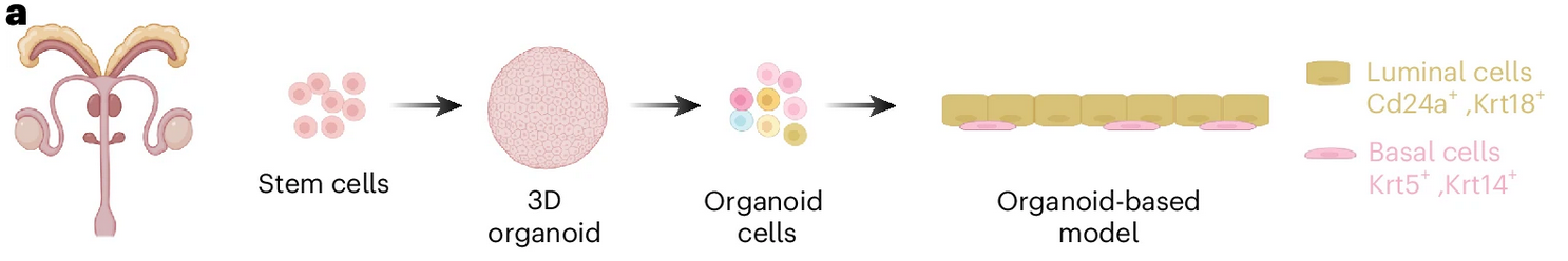
A suitable in vitro infection model should reflect the cellular complexity of the tissue it represents. In case of a prostate model, this should include the main cell types (that is, luminal gland and basal stem cells) present in the prostate epithelium. To set this up, we generated mouse prostate three-dimensional (3D) organoids from C57BL/6 wild-type (WT) mice following a previously published protocol . These organoids were then seeded onto a two-dimensional (2D) surface to provide apical accessibility for subsequent infections. The previously described prostate organoid media (Ctrl) did not achieve a very high degree of differentiation, as observed by the low number of differentiated luminal cells (CD24a+) and the high number of KRT5+ basal cells. Given that activation of the androgen receptor pathway is the primary driver of prostate epithelium development and differentiation, we hypothesized that increasing androgen levels in the medium could improve cellular differentiation. A 10-fold increase of 5α-dihydrotestosterone (DHT) was sufficient to differentiate basal into luminal cells. In contrast, removing DHT enriched the model for basal stem cells. To obtain a more in-depth characterization of the model, we applied single-cell RNA sequencing (scRNA-seq) to the 2D model and 3D organoids grown in both culture conditions (Ctrl and 10nM DHT).